By Phoebe Ingraham Renda

Treg repair responses reveal a delicate balance in healing transplanted hearts. Illustrated by: Phoebe Ingraham Renda.
In a study published in the Journal of Clinical Investigation, researchers from The University of Pittsburgh’s Departments of Surgery and of Immunology and the Thomas E. Starzl Transplantation Institute discovered that a molecule used by immune cells for tissue repair contributes to chronic organ rejection.
Chronic rejection occurs when fibrosis and inflammation affect the blood vessels of a transplanted organ. Even with immunosuppressant medications, fibrosis and associated vasculopathy (diseases affecting blood vessels) continue to damage transplanted organs. As a result, overcoming the 10- to 15-year success barrier in organ transplants remains a challenge.
“In the process of transplanting an organ, there is going to be surgical trauma that needs to be repaired and dealt with by the recipient’s immune system,” says lead author Jordan Warunek, a graduate student in the School of Medicine’s Program in Microbiology and Immunology.

Jordan Warunek, a graduate student in the Program in Microbiology and Immunology at the School of Medicine.
Early injury of transplanted tissue, also called a graft, leads to significant inflammation, Warunek notes. For example, an abundance of proinflammatory macrophages—immune cells responsible for clearing dead cells, debris and pathogens—in grafts is linked to poor outcomes and chronic rejection. Additionally, when a transplant recipient’s T and B cells identify a graft’s alloantigens (molecular markers that the immune system uses to distinguish between self and nonself cells) as nonself, the resulting immune response can cause tissue injury and rejection when not controlled by immunosuppressants. As a result, research efforts have focused on limiting graft injury and inflammation. However, immune-mediated tissue repair mechanisms and their contributions to grafted tissue health are poorly understood.
To identify tissue repair pathways that could serve as therapeutic targets, Warunek; School of Medicine’s Hēth R. Turnquist, professor of surgery and of immunology; Khodor I. Abou-Daya, assistant professor of surgery; and colleagues investigated how losing a known tissue injury signal, interleukin 33 (IL-33), in grafts affects the recipient’s immune cells as they enter the graft. Using a murine heart transplant model, they harnessed the power of single-cell RNA sequencing to define what reparative immune response processes changed when the IL-33 injury signal was absent.
IL-33 in grafts influenced many immune cells but had the greatest effect on regulatory T cells (Tregs). Tregs, recognized for suppressing immune responses, are also thought to aid in tissue repair by producing growth factors in response to IL-33. The team observed that amphiregulin (Areg), a growth factor that supports stromal cells (cells that help build and repair tissues and organs) and drives stem cell proliferation, was significantly upregulated by IL-33 in Tregs.
“We then knocked that repair molecule out of recipient Tregs with the expectation that it would make things worse and embraced the idea that we’d want to augment this pathway,” says Turnquist, who also directs academics and training at the Thomas E. Starzl Transplantation Institute.
To their surprise, the team discovered that long-term graft health improved when Areg signaling was absent from the recipient’s Tregs. They also showed that this repair molecule and its associated reparative cells were contributing to chronic rejection by promoting fibroblast proliferation in grafts.

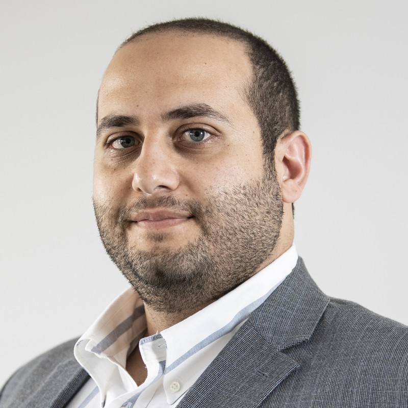
Khodor I. Abou-Daya, assistant professor of surgery, School of Medicine
“What we thought was a good cell is actually dumping molecules leading to fibrosis—which is like cement that is slowly choking the organ out,” says Abou-Daya.
Current research suggests that shortly after cardiac transplantation, IL-33 stimulates reparative macrophages and Tregs to turn on the injury repair process. However, over time, IL-33's role in healing becomes dysregulated—likely due to sustained Treg repair, activated by lingering T and B cell responses to graft alloantigens, or unresolved tissue injury. Dysregulated Treg responses also appear to create fibrotic niches that enable more immune cells to infiltrate the donor tissue and cause localized inflammation. Highlighting a delicate balance in the healing process, their results suggest that turning the Treg repair response off over time could protect against or mitigate fibrotic disease.
The team also cautions that dysregulated IL-33-mediated Treg repair could lead to unexpected side effects from Treg-based cell therapies currently under study—emphasizing the need to better understand Treg repair mechanisms and what drives them.

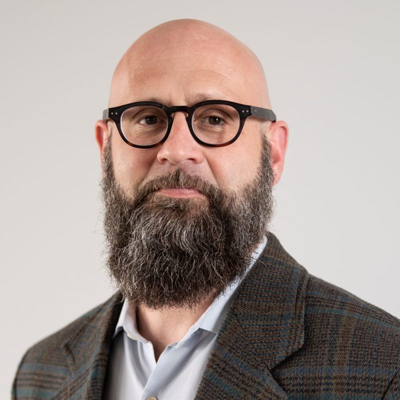
Hēth R. Turnquist, professor of surgery and of immunology, School of Medicine, and director of academics and training at the Thomas E. Starzl Transplantation Institute.
“Instead of just looking for new immunosuppressants, I think one of the next areas in transplant research will be to figure out what repair pathways are good, what repair pathways are detrimental and how and when we target those pathways,” says Turnquist.
This mechanistic understanding of how Treg repair responses generate fibrosis could also help to understand the molecular processes driving T cell-mediated fibrosis in other autoimmune disorders, like systemic sclerosis.
